
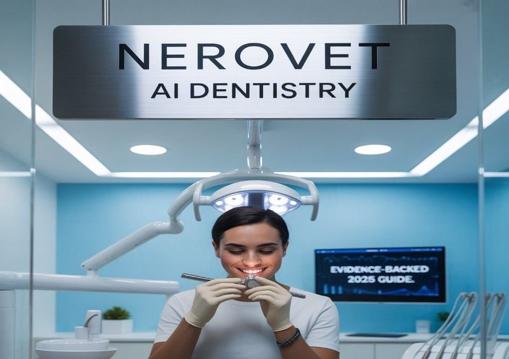
Curious about Nerovet AI dentistry? This guide explains what dental AI actually does in the operatory, how it enhances radiograph interpretation and patient communication, where it falls short, and precisely how to implement it in a clinic without hype.
What Is “Nerovet AI Dentistry”?
Nerovet AI Dentistry is a popular phrase used online to describe AI-assisted dental care — especially systems that analyze radiographs and patient records to support diagnosis and treatment planning. While the label itself isn’t tied to a specific public product, the underlying capability refers to well-established dental AI functions, including highlighting potential caries, quantifying bone levels, identifying calculus, and improving image clarity, which helps dentists make better, faster decisions.
The key idea is to assist the clinician. Artificial intelligence provides consistent templates and assessments, but the final decision-making power remains with the dentist. This clear distinction in responsibilities is crucial for ensuring safe, ethical, and legally sound treatment.
- Dental X-rays include bitewing, periapical, and panoramic views.
- Records: Medical history, periodontal charting, prior treatments.
2) Model Mechanics (Plain English)
Most solutions use deep learning (commonly, convolutional neural networks) trained on expertly annotated images. They learn visual patterns of decay, calculus, and bone level changes. In practice, the system draws subtle boxes/contours, attaches millimeter-level measurements for bones, and flags regions that warrant a closer look.
3) Outputs You’ll See
- Heatmaps or boxes on suspect lesions
- Quantified bone levels to support periodontal staging
- Enhanced images that reduce noise and improve contrast
These visuals standardize what the team sees, reduce misses on early lesions, and make it easier to explain findings to patients.
Real Benefits for Clinics & Patients
- Consistency: Standardized overlays reduce inter-clinician variability in reading bitewings and PAs.
- Earlier detection: Subtle proximal caries and bone changes are easier to flag for review.
- Clarity for patients: Visual evidence improves understanding and typically increases case acceptance.
- Fewer retakes: Enhancement features can clarify underexposed images, cutting friction and saving time.
- Documentation: Structured findings and annotated exports support charting and communication.
Limitations & Risks (Read This)
- Assistive, not autonomous: AI flags “potential” findings; clinical context decides.
- Scope boundaries: Performance is best for the image types and dentitions for which it was trained.
- Dataset bias & drift: New sensors/patient cohorts can shift accuracy if you don’t audit.
- Workflow discipline: Teams need training to interpret overlays and avoid over-treatment.
- Privacy & Consent: Update privacy notices and informed consent to reflect the use of AI.
End-to-End Workflow: From X-ray to Acceptance
- Capture: Acquire quality radiographs; confirm sensor compatibility and exposure presets.
- Analyze: Run AI overlays for caries/bone/calculus; note confidence when available.
- Corroborate: Verify with clinical exam, history, vitality testing when indicated.
- Educate: Show annotated images chairside and explain the risks, options, and prognosis.
- Decide & Document: Final diagnosis and plan by the dentist; export annotated images to the chart.
Nerovet AI Dentistry vs Traditional Methods
Task Traditional Only With AI Assist
Early proximal caries on bitewings. Variable sensitivity across clinicians, Standardized visual cues, and more consistent early flags
Periodontal bone level assessment: Manual estimates; potential variance, Automated mm-level quantification with visuals for patients
Image quality on underexposed films. Possible retakes and time loss. Enhancement reduces noise and clarifies detail.
Implementation Checklist (Copy This)
- Define scope: caries detection, bone measurement, calculus removal, and image enhancement.
- Confirm vendor fit: regulatory status, integration with your PMS/PACS, sensor support.
- Run a 4-week pilot: track detection consistency, retake rate, and case acceptance delta.
- Train the team: reading overlays, communicating findings, and avoiding over-calling.
- Quarterly audits: sample charts; compare AI flags vs clinical outcomes; update SOPs.
- Update documents, including the privacy policy, informed consent, and staff training logs.
- Create patient education assets, including a one-page explainer and annotated examples, to support patient understanding.
FAQs
Is “nerovet ai dentistry” a single product?
No. It’s a popular phrase for AI-assisted dental workflows. Evaluate specific vendors for your indications, integration, and compliance needs.
Will AI replace dentists?
No. AI is assistive. The clinician makes final diagnosis and treatment decisions.
How accurate is dental AI?
Performance is strong for defined tasks (e.g., caries on bitewings) and improves with good image quality, clear indications, and regular audits.
What’s the fastest ROI use case?
Standardizing caries detection and explaining bone loss with mm-level visuals — both tend to improve patient understanding and acceptance.
What about privacy?
Ensure that PHI handling aligns with the requirements of your jurisdiction—update consent forms to reflect the use of AI assistance and data processing.
Next Steps
Ready to implement Nerovet AI dentistry? Start with our downloadable checklist, compare vendors using this scorecard template, and review Dental AI Basics, as well as Patient Communication with AI.